Download Bone and Osteoporosis Measures Data Dictionary
Download Fractures Data Dictionary
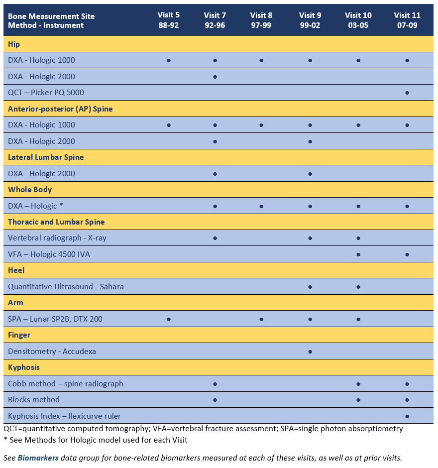
Bone and Osteoporosis Measures began at Visit 5 (1988-92) and continued through Visit 11 (2007-09). This resulted in up to 6 DXA scans of the hip and spine and 5 whole-body scans for each participant over a 20-year period. In addition, several newer bone assessments were conducted including QCT of the hip, vertebral fracture assessment, heel and finger BMD using portable devices, and three measures of kyphosis. Participants from the original cohort were invited to the bone clinic visits when they reached the eligibility age (most were age 45+), and remained eligible as long as they were ambulatory, community dwelling and able to give informed consent.

Bone and Osteoporosis Measures: Summary of Data Available and Methods by Visit
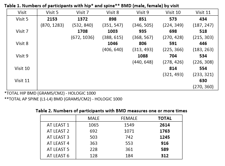
The number of participants with hip and spine scans at each Bone Health visit is shown in Table 1. Table 2 shows the number of participants whose BMD was assessed one or more times. Among all RBS participants, BMD was assessed at least once in 2614, at least twice in 1763, etc.

Bone and Osteoporosis Measures by Visit
Visit 5: Between 1988 and 1991, 2,153 participants, aged 34 to 98 (mean 71.4), had anterior posterior (AP) spine and hip bone mineral density (BMD) measured by dual energy x-ray absorptiometry (DXA) using the Hologic 1000, and ultradistal wrist and midshaft radius BMD measured using single photon absorptiometry (SPA).
Visit 7: Between 1992 and 1996,1,708 participants,aged 30 to 97 (mean 71.0), had repeat DXA of the hip and spine using the Hologic 1000 and first-time DXA scans of the anterior-posterior (AP) spine, the thoracic spine, the lateral lumbar spine and the whole body using the Hologic 2000. Hip axis length was obtained in 947 participants using the Hologic 1000 and repeated in 159 participants using the Hologic 2000.
Visit 8: Between 1997-99, 1,046 participants,aged 37 to 96 (mean 73.6), hadtotal body DXA scans and repeat scans of the hip and lumbar spine BMD using the Hologic 2000.
Visit 9: Between 1998 and 2001, 1,098 participants, aged 42 to 99 (mean 73.7), had repeat DXA of the hip and spine using the Hologic 1000, repeat DXA scans of the anterior-posterior (AP) spine, lumbar spine and whole body using Hologic 2000,and the first heel ultrasound using Hologic Sahara. Lateral x-rays of the thoracic and lumbar spine were repeated.
Visit 10: Between 2003 and 2005, 814 participants, aged 43 to 99 (mean 73.2), had repeat measures of BMD (hip, spine, radius and whole body) and heel ultrasound using the same instruments as Visit 9. Vertebral Fracture Assessment (VFA) using Hologic Delphi DXA was measured for the first time. Lateral spine x-rays were repeated.
Visit 11: Between 2007 and 2009, 630 participants, aged 50 to 104 (mean 75.7), had repeat measures of BMD (hip, spine, radius and whole body), lateral spine x-rays, VFA (vertebral fracture assessment) and heel ultrasound using the same instruments as prior visits. QCT (quantitative computed tomography) of the femoral neck of the hip was obtained for the first and only time.
Bone and Osteoporosis Measure Methods
MEASURES OBTAINED BY EACH METHOD
Detailed descriptions of these methods can be found in the cited references. For unpublished data, more detailed descriptions of data collection are provided, along with links to measurement protocols in select cases.
DXA Scans
Whole body scans (Hologic 2000 – V7, V8, V9; Hologic 4500 – V10; Hologic Discovery W – V11) included assessment of total and regional BMD (bone mineral density). Body composition results (lean mass, fat mass, bone mineral content) from the whole body scans are included in the Physical Characteristics data group.
Hip scans (Hologic 1000, Hologic 2000) assessed the femoral neck, trochanter, intertrochanter, Ward’s triangle and total hip. (2) Hip axis length was determined at Visit 7. (3)
Anterior-posterior (AP) spine scans (Hologic 1000, Hologic 2000) included assessments of the AP spine at L1, L2, L3, L4 and the total AP spine (L1-L4).(4)
Lateral lumbar spine scans (Hologic 2000) included lateral scans at mid L2, mid L3, and mid L4, as well as at L2, L3, L4, and total measures for both areas. (2, 4)
VFA – Vertebral Fracture Assessment (Hologic 4500) assessed vertebral fractures of the thoracic and lumbar spine from lateral spine scans at Visits 10 and 11 using DXA and IVA (instant vertebral assessment) software provided by Hologic. Low-dose, single-energy acquisition mode was performed during short breath hold. The scan time averaged 10 seconds with <10 μSv radiation dose exposure. The fan-beam DXA images for quantitative morphometric assessment of spinal fractures were read independently of spine radiographs at the same visit. IVA results were read for fractures by skeletal radiologists at UCSF, including Harry K Genant, MD. These scans also provided measures of scoliosis, osteophytes, coronary aortic calcification (5) and aortic aneurysm. [Note: As of 2019, the VFA data have not been published.]
Vertebral x-rays at Visits 7, 9 and 10evaluated morphometric fractures of the thoracic and lumbar spine, disk narrowing, osteophytes, DISH (diffuse idiopathic skeletal hyperostosis), juvenile and senile kyphosis, scoliosis, congenital malformations, surgical changes and Paget’s disease.At Visit 7, morphometric spine fractures were evaluated by a single musculoskeletal radiologist. Several months after the first reading, 60 radiographs were re-examined by the same radiologist (without any knowledge of his first reading), with only two disagreements (Kappa: 0.82; i.e., proportion agreement: 96%). (4, 6, 7)
Peripheral Scans by SPA (single photon absorptiometry) assessed bone mineral content (BMC) and BMD of the ultradistal radius (10 slices) and the midshaft radius (4 slices) at Visit 5 using the Lunar SP2B (2, 7). At Visits 8, 9 and 10, the distal radius, radius, ulna and nROI (region of interest) were evaluated using the DTX-200 Osteometer. At the start of Visit 8 (03-06-97 to 11-24-98), the DTX-200 used software version 1.54. Version 1.63 was used for the remainder of Visit 8 and for Visits 9 and 10. [Note: As of 2019, the peripheral scan data from Visits 8, 9 and 10 have not been published.]
Quantitative Ultrasound (Sahara Clinical Bone Sonometer) of the calcaneous (heel bone) was conducted at Visits 9 and 10. The Sahara measures speed of sound (SOS in m/s) and broadband ultrasonic attenuation (BUA in dub/mHz) of an ultrasound beam passed through the calcaneous, and combines these results linearly to obtain the Quantitative Ultrasound Index (QUI). The output is also expressed as a T-score and as an ultrasound estimate of the BMD (g/cm) of the calcaneous as measured by DXA. [Note: As of 2019, these data have not been published.]
Densitometry (Accudexa) of the finger was performed at Visit 9 using Accudexa software version 1.30. [Note: As of 2019, these data have not been published.]
QCT (quantitative computed tomography) (Picker PQ-5000) of the femoral neck of the hip was obtained at Visit 11 (See Black DM et al, JBMR, 23(8): 1326-1333, 2008 for a detailed description of methods used). The QCT scans were performed at Poway Imaging Center (Poway, CA). [Note: As of 2019, these data have not been published.]
Kyphosis was assessed by 3 methods: a modified Cobb method (Visit 7, 10 and 11) (8), the “blocks” method (Visit 7, 10 and 11) (9) and by a Kyphosis Index based on flexicurve (Athena) measurements of the spine (Visit 11) (10). The flexicurve measurements were obtained with participants standing with their normal posture and during maximal achievable erectness (see Kyphosis Index Measurement for a diagram of measurement variables).
Fractures
Download Fractures Data Dictionary
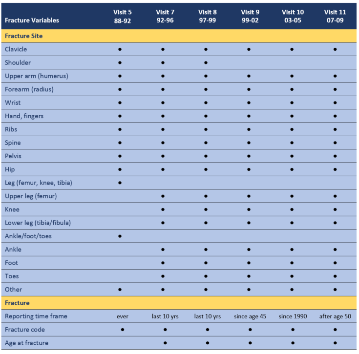
Fracture history was obtained at each research clinic Visit and via multiple Mailers using self-administered questionnaires. Participants were queried by fracture site for fracture (yes/no), year of fracture, age at fracture and cause of fracture (chosen from a list of 9 codes encompassing severe trauma, 3 types of falls, spontaneous fracture, and local bone disease). The time frame for the fracture history questions varied by Visit as indicated in the summary table below. Clinic nurses reviewed the fracture history questionnaire with participants. History of reported clinical fractures was validated in a subset of 36%, with validation of 71% by medical chart review (1). Vertebral fractures identified by spine scans are included in the section on Bone and Osteoporosis Measures.

References
- Jassal SK, von Muhlen D, Barrett-Connor E. Measures of renal function, BMD, bone loss, and osteoporotic fracture in older adults: the Rancho Bernardo study. J Bone Miner Res. 2007;22(2):203-10. doi: 10.1359/jbmr.061014. PubMed PMID: 17059370; PMCID: PMC2895929.
- Edelstein SL, Barrett-Connor E. Relation between body size and bone mineral density in elderly men and women. Am J Epidemiol. 1993;138(3):160-9. PubMed PMID: 8356959.
- Clark P, Tesoriero LJ, Morton DJ, Talavera JO, Karlamangla A, Schneider DL, Wooten WJ, Barrett-Connor E. Hip axis length variation: its correlation with anthropometric measurements in women from three ethnic groups. Osteoporos Int. 2008;19(9):1301-6. doi: 10.1007/s00198-008-0572-8. PubMed PMID: 18301856; PMCID: PMC2701735.
- Schneider DL, Bettencourt R, Barrett-Connor E. Clinical utility of spine bone density in elderly women. J Clin Densitom. 2006;9(3):255-60. doi: 10.1016/j.jocd.2006.04.116. PubMed PMID: 16931341; PMCID: PMC2642644.
- Schousboe JT, Claflin D, Barrett-Connor E. Association of coronary aortic calcium with abdominal aortic calcium detected on lateral dual energy x-ray absorptiometry spine images. Am J Cardiol. 2009;104(3):299-304. doi: 10.1016/j.amjcard.2009.03.041. PubMed PMID: 19616658; PMCID: PMC2763778.
- Trone DW, Kritz-Silverstein D, von Muhlen DG, Wingard DL, Barrett-Connor E. Is radiographic vertebral fracture a risk factor for mortality? Am J Epidemiol. 2007;166(10):1191-7. doi: 10.1093/aje/kwm206. PubMed PMID: 17709329.
- Hongsdusit N, von Muhlen D, Barrett-Connor E. A comparison between peripheral BMD and central BMD measurements in the prediction of spine fractures in men. Osteoporos Int. 2006;17(6):872-7. doi: 10.1007/s00198-005-0061-2. PubMed PMID: 16525761.
- Schneider DL, von Muhlen D, Barrett-Connor E, Sartoris DJ. Kyphosis does not equal vertebral fractures: the Rancho Bernardo study. J Rheumatol. 2004;31(4):747-52. PubMed PMID: 15088302.
- Kado DM, Huang MH, Karlamangla AS, Barrett-Connor E, Greendale GA. Hyperkyphotic posture predicts mortality in older community-dwelling men and women: a prospective study. J Am Geriatr Soc. 2004;52(10):1662-7. doi: 10.1111/j.1532-5415.2004.52458.x. PubMed PMID: 15450042.
- Wankie C, Kritz-Silverstein D, Barrett-Connor E, Kado DM. Kyphosis and Sleep Characteristics in Older Persons: The Rancho Bernardo Study. J Sleep Disord Manag. 2015;1(1). PubMed PMID: 28480455; PMCID: PMC5419044.
